
Biochemistry - ENZYMES
Про матеріал
MCAT based biochemistry lesson Presentation on Enzymes and their properties, functions and fundamentals.
M C AT B I O C H E M I S T R Y · L E S S O N 1
Enzymes: the body’s
catalysts
Structure, kinetics, inhibition and regulation, plus the graphs the MCAT loves
Dr. Paramvir Singh

F O U N D AT I O N S
Enzymes are biological catalysts
• A catalyst speeds up a reaction without being used up
• Nearly all enzymes are proteins; a few are RNA molecules called
ribozymes
• Each enzyme is specific to one substrate or one type of reaction
• Enzymes change the rate, not the final balance of a reaction
• Cells need them: uncatalyzed reactions at 37°C are far too slow
Speed-up
10
⁶
and up
Typical rate increase; some
enzymes exceed this by many
orders of magnitude
Enzymes
Speed-up
10
⁶
=
1000000 =
1 millon

Enzymes and their Sites

What an enzyme actually does
Lowers activation energy
Stabilizes the transition state, so more molecules clear the barrier
each second.
Changes nothing else
Δ
G, Keq and the direction of the reaction stay the same.
Enzymes only get you to equilibrium faster.
Comes out unchanged
Reusable and specific. Nearly all are proteins; catalytic RNAs
(ribozymes) are the rare exception.
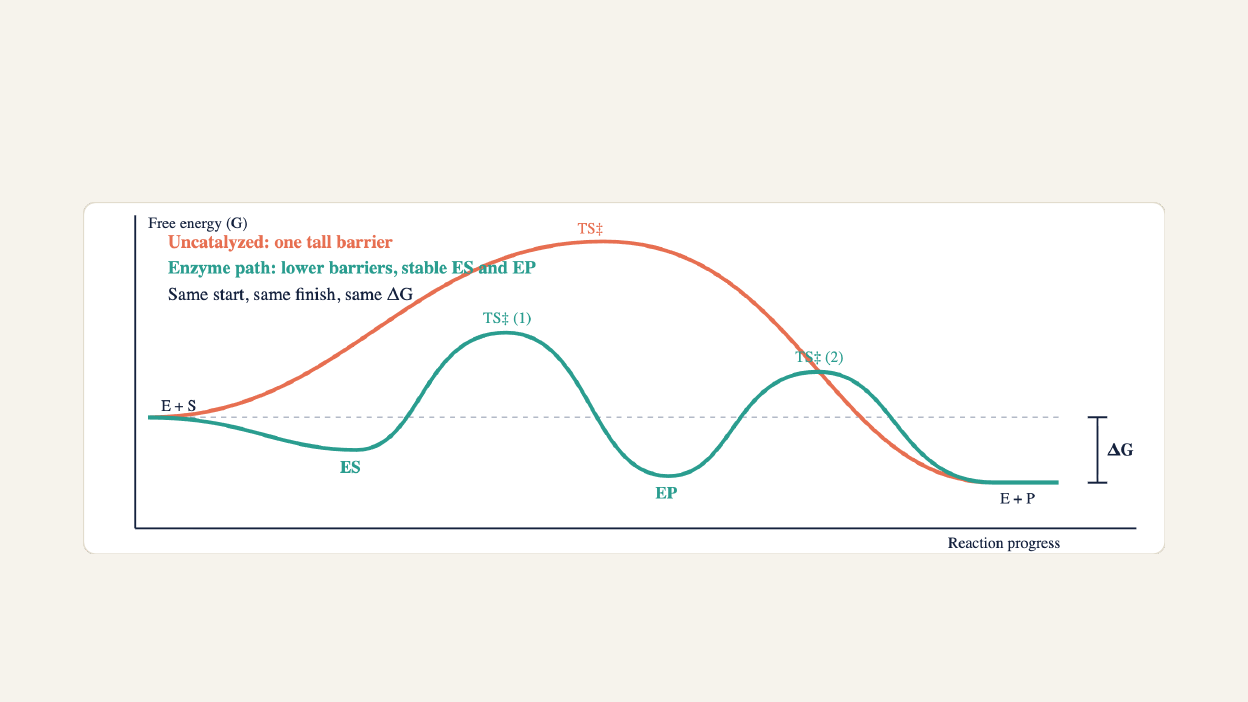
The reaction coordinate, step by step

Anatomy of an enzyme
E + S
ES
→
EP
→
E + P
Apoenzyme (protein) + cofactor =
holoenzyme
Active site and induced fit
A pocket of specific amino acid side chains. The substrate
binds noncovalently and the enzyme reshapes around it
(induced fit), rather than a rigid lock and key.
Cofactors
Inorganic ions such as Zn
²⁺
, Mg
²⁺
and Fe
²⁺
. Often loosely
bound, and needed for activity (carbonic anhydrase uses
Zn
²⁺
).
Coenzymes
Small organic carriers, mostly vitamin-derived: NAD
⁺
(B3),
FAD (B2), CoA (B5), TPP (B1), PLP (B6), biotin, THF (folate),
B12.
Prosthetic groups
Cofactors bound tightly or covalently and permanently, such
as the heme in cytochromes. Strong acid is needed to pull
them off.

Lock and key vs induced fit
Lock and key
Older model: the site is pre-shaped and only the matching substrate
fits. It explains specificity but not catalysis.
Induced fit (MCAT*)
Binding reshapes the site, aligns catalytic residues and strains the
substrate toward the transition state.

Vitamins become coenzymes
Vitamin
Coenzyme
Job
B1 thiamine
TPP (Thiamine Pyrophosphate)
Decarboxylation, aldehyde transfer (PDH)
B2 riboflavin
FAD, FMN (Flavin Adenine Dinucleotide,
Redox, carries electrons
B3 niacin
NAD
⁺
, NADP
⁺
(Nicotinamide Adenine
Dinucleotide Phosphate)
Redox, carries hydride
B5 pantothenate
COA (Coenzyme A)
Acyl group transfer
B6 pyridoxine
PLP (Pyridoxal Phosphat)
Amino group transfer (transamination)
B7 biotin
Biocytin
Carboxylation (uses ATP)
B9 folate
THF (Tetrahydrofolate)
One-carbon transfer
B12 cobalamin
Methyl- and adenosylcobalamin
Methyl transfer, isomerization
Coenzymes are consumed and regenerated, so they act as co-substrates. Deficiency stalls the matching enzyme, for example thiamine
deficiency and beriberi.

The six enzyme classes (OTHLIL)
Class
What it does
Examples
1 Oxidoreductases
Redox: move electrons or H
Lactate dehydrogenase, cytochrome
oxidase
2 Transferases
Move a functional group
Kinases, ALT/AST (transaminases)
3 Hydrolases
Break bonds using water
Proteases, lipases, phosphatases
4 Lyases
Break or form bonds without water or ATP,
making a double bond
Aldolase, fumarase, decarboxylases
5 Isomerases
Rearrange within one molecule
Triose phosphate isomerase, mutases
6 Ligases
Join two molecules, powered by ATP
DNA ligase, pyruvate carboxylase
Name tricks:
synthase (no ATP) is a lyase, synthetase (uses ATP) is a ligase. Kinase moves phosphate using ATP, phosphatase
removes it with water.

Michaelis–Menten kinetics
v
₀
= Vmax[S] / (Km + [S])
Vmax and kcat
Vmax is the speed when every enzyme is saturated. It scales with
enzyme amount: kcat = Vmax / [E]total.
Km
The [S] giving
½
Vmax. Low Km means high affinity. Below Km the
rate is first order in [S]; far above Km it is zero order.
Allosteric enzymes
Cooperative, so the curve is sigmoidal, not hyperbolic.

Comparing two enzymes: same Vmax, different Km
Enzyme A: Km 2 mM
Half-saturated at a tiny [S]. High affinity.
Enzyme B: Km 10 mM
Needs five times more substrate to reach
½
Vmax. Lower affinity.
Read the graph
Find
½
Vmax on the y-axis, go across to the
curve, then drop down to read Km.

The same two enzymes on a Lineweaver–Burk plot
1/v = (Km/Vmax)(1/[S]) + 1/Vmax
Intercepts
y-intercept = 1/Vmax. x-intercept =
−
1/Km. Slope =
Km/Vmax.
Why it helps
A straight line is easy to extrapolate and compare.
Errors at low [S] get amplified, though.

Reading inhibition on a Lineweaver–Burk plot
Competitive
Binds the active site, mimics substrate.
Vmax same, Km up. High [S]
outcompetes it.
Noncompetitive
Binds an allosteric site on E or ES. Vmax
down, Km same. Reversible, and more
[S] cannot fix it.
Uncompetitive
Binds only the ES complex. Both Vmax
and Km go down.
Gray = no inhibitor, orange = inhibitor. Irreversible inhibitors bind covalently and permanently, and are a separate category.

The same inhibitors on Michaelis–Menten curves
Competitive
The curve shifts right and still reaches the
same ceiling at high [S].
Noncompetitive
Some enzyme is out of action, so the
ceiling drops. Half-max sits at the same
[S].
Uncompetitive
Trapping ES lowers the ceiling and also
pulls Km left.
Gray = no inhibitor, orange = inhibitor. Dashed lines mark Vmax and Km.

How the cell turns enzymes up and down
Allosteric
Effectors bind a separate site and shift the
enzyme between low-affinity (T) and high-
affinity (R) states. Gives sigmoidal curves.
Covalent modification
Kinases add phosphate to Ser, Thr or Tyr,
phosphatases remove it. Reversible, and
can switch an enzyme on or off.
Zymogens
Inactive precursors cut once to activate,
irreversibly: trypsinogen to trypsin,
pepsinogen to pepsin.
Feedback inhibition
The end product inhibits an early enzyme
of its own pathway, so the cell never
overproduces.
Temperature
Rate climbs, peaks near 37°C for human
enzymes, then collapses as the protein
denatures. “Doubles per 10°C” holds only
well below the peak.
pH optimum
Pepsin works near pH 2 in the stomach,
trypsin near pH 8 in the small intestine.
Wrong pH changes charges and shape.

Allosteric enzymes: the sigmoidal curve
T state and R state
Tense is low affinity, relaxed is high affinity. One bound
substrate nudges the other subunits toward R:
cooperativity.
Effectors
Positive effectors shift the curve left. Negative effectors
shift it right.
Classic example
Hemoglobin binding O
₂
follows the same S-shaped logic.

Temperature and pH optima
Temperature: activity rises, peaks, then collapses because the protein unfolds. pH: charges on active-site residues shift, so each enzyme works best
in its home environment.

Isoenzymes and diagnostic enzymes
Isoenzymes catalyze the same reaction in different tissues but differ in kinetics and regulation. Damaged cells leak them into
blood, so their pattern points to the organ.
Marker
Points to
Key fact
LDH1 (H
₄
) vs LDH5 (M
₄
)
Heart vs liver and muscle
LDH is a tetramer of H and M subunits,
giving five isoenzymes
CK-MB
Myocardial infarction
Rises in 4–8 h, peaks about 24 h, normal
by 48–72 h
Cardiac troponin
Myocardial infarction
Rises in 4–6 h, stays up 3–10 days, the
gold standard
ALT and AST
Liver injury
ALT is the more liver-specific one
Amylase, lipase
Pancreatitis
Pancreatic enzymes leaking into blood

Test yourself
1. A drug raises apparent Km but leaves
Vmax unchanged. Where do the
Lineweaver–Burk lines cross, and how
can you overcome it?
Competitive. On the y-axis. Add more
substrate.
2. An inhibitor lowers Vmax and leaves
Km alone. What is its type, and where
do the lines cross?
Noncompetitive. On the x-axis, at
−
1/
Km.
3. A patient arrives five days after chest
pain. Which marker is more useful, CK-
MB or troponin?
Troponin. It stays elevated 3–10
days; CK-MB is normal by 48–72
hours.
Lesson 1 in one line:
enzymes speed things up without shifting equilibrium. Read Km and Vmax from the graph, and
match every inhibitor to its Lineweaver–Burk signature.
 Зареєструватись за допомогою Google