Презентація "Серце, будова та його функції"


Heart. Performed: Viktor Van Art
1. Anatomy Heart - 32. Atrium and ventricle - 43. Blood pumps – 5-64. Conduction system – 7 5. Blood circulation – 8 6. Heart situated – 9 7. The largest part – 10 8. Valves – 11 9. The heart wall – 12-1310. Rytm heart – 14 11. Сontinue – 15 12. Еnd of rhytm – 16 13. Аdditionally. List
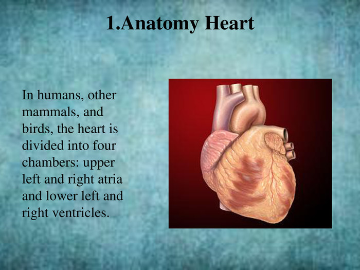
In humans, other mammals, and birds, the heart is divided into four chambers: upper left and right atria and lower left and right ventricles.1. Anatomy Heart
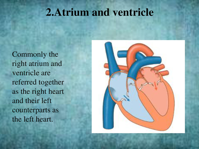
Commonly the right atrium and ventricle are referred together as the right heart and their left counterparts as the left heart.2. Atrium and ventricle
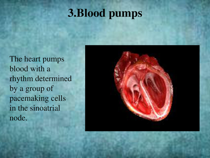
The heart pumps blood with a rhythm determined by a group of pacemaking cells in the sinoatrial node.3. Blood pumps
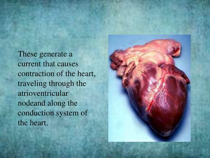
These generate a current that causes contraction of the heart, traveling through the atrioventricular nodeand along the conduction system of the heart.
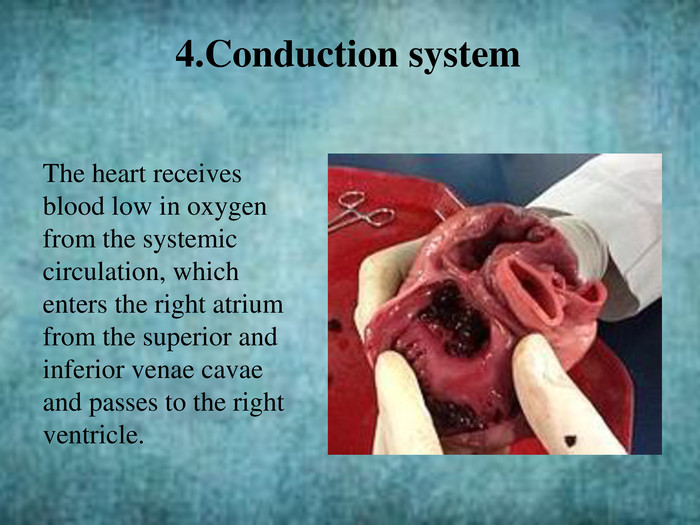
4. Conduction system. The heart receives blood low in oxygen from the systemic circulation, which enters the right atrium from the superior and inferior venae cavae and passes to the right ventricle.
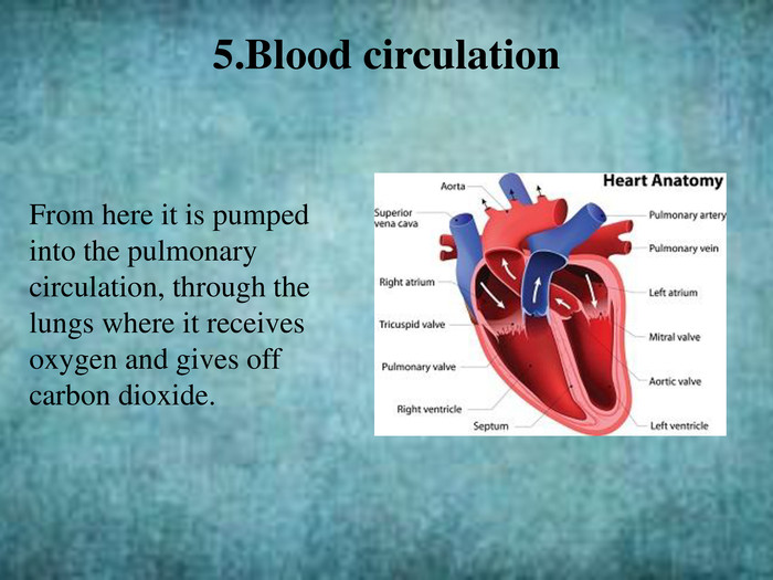
From here it is pumped into the pulmonary circulation, through the lungs where it receives oxygen and gives off carbon dioxide.5. Blood circulation
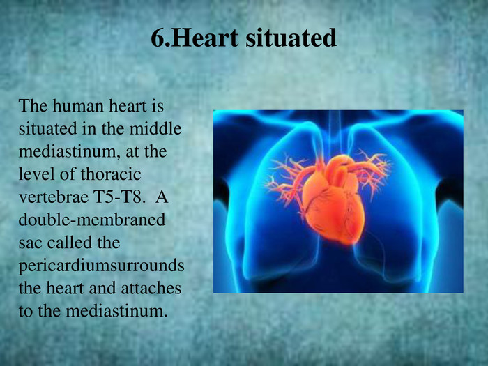
6. Heart situated. The human heart is situated in the middle mediastinum, at the level of thoracic vertebrae T5-T8. A double-membraned sac called the pericardiumsurrounds the heart and attaches to the mediastinum.
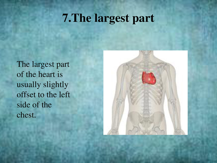
7. The largest part. The largest part of the heart is usually slightly offset to the left side of the chest.
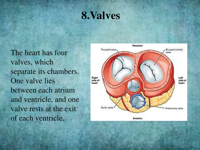
8. Valves. The heart has four valves, which separate its chambers. One valve lies between each atrium and ventricle, and one valve rests at the exit of each ventricle.
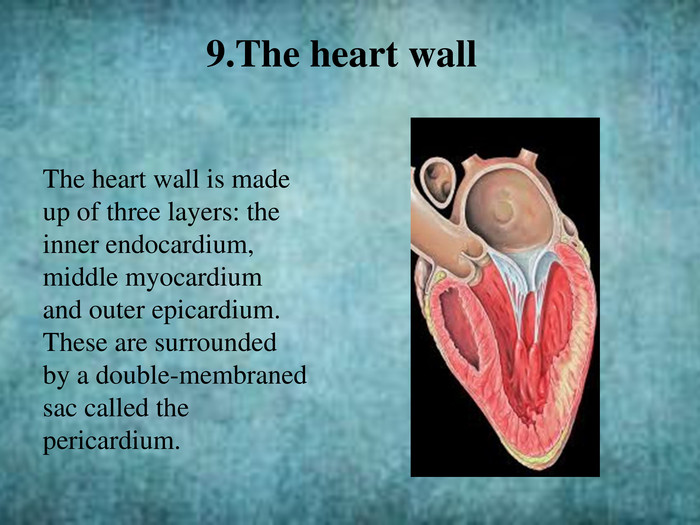
The heart wall is made up of three layers: the inner endocardium, middle myocardium and outer epicardium. These are surrounded by a double-membraned sac called the pericardium.9. The heart wall

Аnd is felt to be on the left because the left heart is stronger and larger, since it pumps to all body parts.
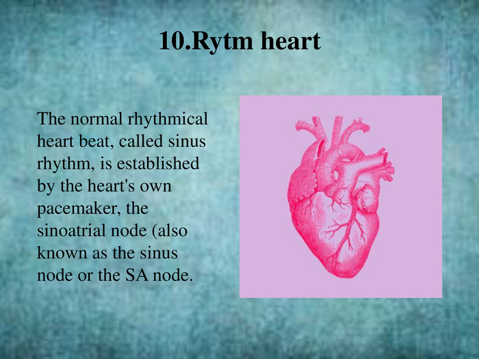
10. Rytm heart. The normal rhythmical heart beat, called sinus rhythm, is established by the heart's own pacemaker, the sinoatrial node (also known as the sinus node or the SA node.
Here an electrical signal is created that travels through the heart, causing the heart muscle to contract. 11. Сontinue
12. Еnd of rhytm. The sinoatrial node is found in the upper part of the right atrium near to the junction with the superior vena cava.

did you think this is the end???P. S. I tried to make this presentation, I hope it came out well, thanks for spending your time and checking my work. Best regards Nick


про публікацію авторської розробки
Додати розробку