Статья Гемангіоми бластоми.клінічні випадки
ГЕМАГІОБЛАСТОМА ГОЛОВНОГО МОЗКУ
Данилова Інеса Віталіївна
Магістр житомир медичний інститут
Кафедра охрони здоров'я


Анотація:
На сьогодні Гемангіобластоми – це достатньо рідкісні доброякісні судинні пухлини головного мозку.
У статті були описані клінічні випадки з діагнозом Гемангіостоми.
У таких пацієнтах наблюдався ,сильний головний біль , з вираженою мігренню ,спазми гол мозку судин, інтосикація ,прушення розмови та парез нижніх та вер. кінцівок.
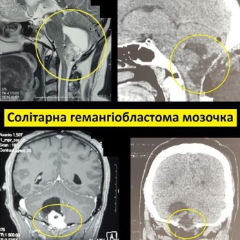
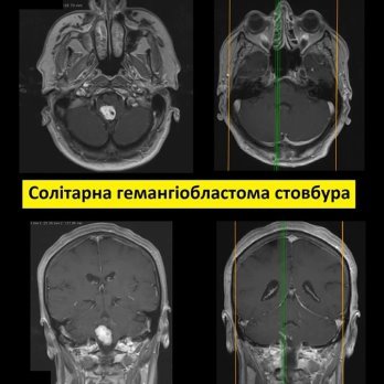
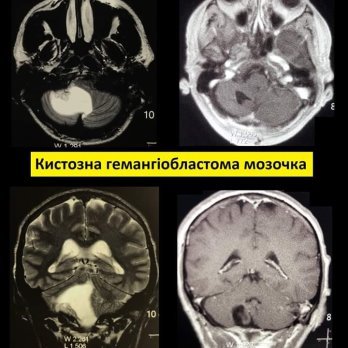
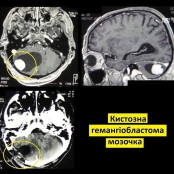
На КАТ та МРТ спостерігалась кістозна гемангіопластома мозочка( опухоль),з корововиливом в деяких випадках.
Аналіз крові розгорнутий, та додаткові обстеження .Анамнез життя.
За останні півроку я в своїй практиці зустрілася 4 пацієнтами з таким діагнозом і сьогодні хочу презентувати свої результати їхнього лікування,де було проведено обстеження в Київської лікарні швидкої допомоги м. Києва .
Паціенти такі були однакового віку від55-68 років,однакового типу захворюваності продовж життя.
В анамнезі таких паціентів були інші паталогії гіпертонія ,гостра хвороба серця, серцева недостатність та інсульти.
Після таких випадків хвороби -це мало статися.
Таки випадки були цікавими та одностороніми.
Загально було проведено обстежиння лікарем -интерном магстратури Житомирського медичного інституту ,та іншими лікарями нейрохірургчнього віділенні.
Приймальне відділення та нейро відділенні невідкладної допомоги .
Актуальність теми:
Гемангіобластома головного мозку може бути самостійним захворюванням (70% випадків), або бути проявом рідкісної генетичної хвороби - фон Гіппель-Ліндау (30% випадків).
Хвороба фон Гіппель-Ліндау – це мутація гена VHL, що розташований на 3 хромосомі. В наслідок цієї мутації людина має підвищену схильність до утворення пухлин, зокрема в головному та спинному мозку, сітківці, печінці, нирках, підшлунковій залозі.
Проаналізувавши на сьгодні , що основним і фактично єдиним методом лікування гемангіобластоми є її хірургічне видалення. Складається пухлина з великої кількості судин, що мають тонку ламку стінку. Особливістю видалення гемангіобластоми є необхідність першочергового від’єднання пухлини від живлячих судин і лише потім видалення пухлини одним блоком. Цим гемангіобластоми чимось нагадують АВМ (артеріо-венозні мальформації). Якщо намагатися видаляти пухлину частинами, то це може викликати жахливу кровотечу.
Коли на МРТ головного мозку виявляють гемангіобластому, то дуже важливим є першочергове виконання МРТ всього спинного мозку, оскільки не рідко гемангіобластом може бути кілька.
Висновок:
У всіх 4випадках, що оперувалися в нас вдалося досягти тотального видалення пухлини та уникнути будь якого неврологічного дефіциту.
Отже: да оперція була вдалой та досягнула успіпів в хірургічному лікуванні,без рецедивів.
Annotation: Today, hemangioblastomas are quite rare benign vascular tumors of the brain. The article described clinical cases with a diagnosis of hemangiostoma. In such patients there was a severe headache, with severe migraine, spasms of the cerebral vessels, intoxication, speech disturbances and paresis of the lower and vertebrae. limbs. Cystic hemangioplastoma of the cerebellum (tumor) was observed on CAT and MRI, with hemorrhage in some cases. Blood test is detailed, and additional examinations. Life history. Over the past six months, I have met 4 patients with this diagnosis in my practice and today I want to present my results of their treatment, where an examination was conducted at the Kyiv Ambulance Hospital in Kyiv. Such patients were of the same age from 55-68 years, the same type of morbidity throughout life. Such patients had a history of other pathologies of hypertension, acute heart disease, heart failure and stroke. After such cases, the disease was about to happen. Such cases were interesting and one-sided. In general, an examination was conducted by an intern master of the Zhytomyr Medical Institute, and other doctors of the neurosurgical department. Admission department and neuro department of emergency care. Actuality of theme: Hemangioblastoma of the brain can be an independent disease (70% of cases), or be a manifestation of a rare genetic disease - von Hippel-Lindau (30% of cases). Von Hippel-Lindau disease is a mutation in the VHL gene, which is located on chromosome 3. As a result of this mutation, a person has an increased tendency to form tumors, in particular in the brain and spinal cord, retina, liver, kidneys, pancreas. Analyzing today that the main and in fact the only method of treatment of hemangioblastoma is its surgical removal. The tumor consists of a large number of vessels with a thin fragile wall. The peculiarity of hemangioblastoma removal is the need to first separate the tumor from the feeding vessels and only then remove the tumor in one block. This hemangioblastoma somewhat resembles AVM (arterio-venous malformations). If you try to remove the tumor in parts, it can cause terrible bleeding. When hemangioblastoma is detected on MRI of the brain, it is very important to perform primary MRI of the entire spinal cord, as it is not uncommon for there to be several hemangioblasts. Conclusion: In all 4 cases operated on, we managed to achieve total removal of the tumor and avoid any neurological deficit. So: yes, the operation was successful and achieved success in surgical treatment, without recurrence.
Література
1. Vitovskiy RM, Beshliaha VM. Features of diagnostics and surgical treatment of primary cardiac tumors. Medycni initsiatyvy. 2014;3(5). (in Ukr.).
2. Abad C, de Varona S, Limeres MA. et al. Resection of a left atrial hemangioma: report of a case and overview of the literature on resected cardiac hemangiomas. Tex Heart Inst. J. 2008;35:69–72.
3. Esmaeilzadeh M, Jalalian R, Maleki M. et al. Cardiac cavernous hemangioma. Eur J Echocardiography. 2007;8(6):487–489.
4. Han Y, Chen X, Wang X. et al. Cardiac capillary hemangioma: a case report and brief review of the literature. J Clin Ultrasound. 2014;42:53–56


про публікацію авторської розробки
Додати розробку
